People who live with depression have low blood levels of a specific molecule, new medical research has revealed. It’s called acetyl-L-carnitine, and those with particularly severe, treatment-resistant or childhood onset depression were found to have the lowest levels.
Naturally produced by the body, acetyl-L-carnitine plays a crucial
role in metabolising fat and the production of energy. It’s also widely
available as a dietary supplement – not some strange and esoteric thing.
Now
researchers from multiple institutions have found a link to depression,
noticing a clear correlation between the condition and noticeably low
levels of acetyl-L-carnitine.
In recent years, more and more
evidence has been building to suggest this link. Since at least 1991,
medical researchers have been aware of acetyl-L-carnitine’s potential to
treat depression, particularly in geriatric and comorbid patients, with the substance showing greater efficacy than a placebo.
More recently, Carla Nasca of the Rockefeller University led a study on rodents,
which found that acetyl-L-carnitine had a fast-acting antidepressant
effect on rats, kicking into effect in just a few days, rather than the
weeks it takes for drugs like SSRIs.
Now Nasca and colleagues have conducted a study on human patients to see if there’s a basis for a similar trial in people.
“It’s the number one reason for absenteeism at work, and one of
the leading causes of suicide. Worse, current pharmacological
treatments are effective for only about 50 percent of the people for
whom they’re prescribed. And they have numerous side effects, often
decreasing long term compliance.”
The research team recruited 71
patients with a diagnosis of depression. These were men and women, aged
between 20 and 70. They also recruited 45 demographically matched
healthy controls.
The patients had to fill out a detailed
questionnaire, undergo a clinical assessment and medical history, and
give a blood sample. Of the patients with depression, 28 had moderate
depression and 43 had severe depression at the time of the study.
When
compared to the age- and sex-matched healthy controls, the patients
with depression had substantially lower levels of acetyl-L-carnitine.
Those
with the most severe depression had the lowest levels. This included
patients whose depression had resisted antidepressant drugs, those with
early onset, and those who had experienced childhood abuse, neglect,
poverty or violence.
These patients constitute around 25-30
percent of all people suffering depression, and are the most in need of
help, the researchers said.
But there are a few steps to be done before acetyl-L-carnitine
supplements can be approved as a treatment. In particular, clinical
trials on human patients with depression, since, as we know, results from rodent models can’t always be replicated in humans.
The
researchers also don’t know the reason for the correlation, or the
effect it has. The rat research suggests that acetyl-L-carnitine plays a
role in the brain, preventing the excessive firing of excitatory
neurons, but this will need to be explored further as well.
“We’ve identified an important new biomarker of major depression disorder,” Rasgon said.
“We
didn’t test whether supplementing with that substance could actually
improve patients’ symptoms. What’s the appropriate dose, frequency,
duration? We need to answer many questions before proceeding with
recommendations, yet. This is the first step toward developing that
knowledge, which will require large-scale, carefully controlled clinical
trials.”
And we’ll be eagerly awaiting the results of those trials.
Meanwhile, the team’s research can be found in the journal PNAS.
Here in Canada, the government agency which is responsible for licensing natural health products is the NNHPD – Natural and Non-Prescription Health Products Directorate.
The regulatory approval process to get natural health products approved can be challenging, frustrating and take a long time: some of our formulations have taken over 2 years before they were finally approved!
Nevertheless, we feel that this regulatory framework ultimately makes for a better regulated market which ultimately helps consumers.
Our company, Robert Lamberton Consulting and our various brands: Cutting Edge Naturals, SmartBrain Formulations and OPN – Optimum Performance Nutrition follow these guidelines.
You may be swallowing billions of tiny plastic particles while sipping a cup of freshly brewed gourmet tea, a new study from McGill University in Montreal suggests.
Many
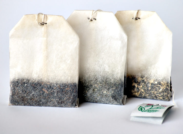
fancier teas now come in “silken” bags instead of paper. Some of them
are pyramid-shaped, which is billed as a way to make room for the large
leaves in premium teas to expand.
Nathalie Tufenkji, a
professor of chemical engineering at the Montreal university, was
surprised to find one such bag in the tea she ordered from a coffee shop
one morning.
It looked like plastic, she recalled. “I said, ‘Oh God, I’m sure if it’s plastic it’s, like, breaking down into the tea.'”
So when she got into the lab, she asked her graduate student, Laura Hernandez, to go out and buy a bunch from different brands.
Sure
enough, Hernandez’s lab tests showed that when steeped in hot water,
the tea bags released microplastic and even smaller nanoplastic
particles — and not just the hundreds or thousands Tufenkji had been
expecting.
“We were shocked when we saw billions of particles in a single cup of tea,” she said.
One
cup from a single tea bag could contain 11.6 billion microplastic and
3.1 billion nanoplastic particles, the researchers estimated from their
results, published Wednesday in the journal Environmental Science & Technology.
The
bits are so tiny — on average, the size of grains of dust or pollen
— that the amount in one cup is about 16 micrograms or one-sixtieth of a
milligram of plastic.
But that’s still much more than has been found in other foods and beverages, including tap and bottled water, beer, honey, fish and shellfish, chicken
and salt. Tufekji said that’s partly because her study included and
counted smaller particles than most other studies. But it’s also
because, for most foods and beverages, the plastic is an accidental
contaminant. With tea “you’re literally adding plastic into the
beverage.”
That may sound unappetizing, but is it a health risk?
The
Tea and Herbal Association of Canada told CBC News in a statement that
the materials used in the tea bags in the study, PET (polyethylene
terephthalate, found in plastic drink bottles) and nylon (used in many
food bags and pouches), have been deemed safe for use in contact with
hot food and beverages. It added there is no evidence the microparticles
pose a risk to human health.
But in general, there isn’t much known on the impact of microplastics on human health.
“There’s
really no research. But this really points to the need to do those
studies,” Tufenkji said. “Think of people who drink one or two or three
cups of tea a day, every day.”
The
researchers counted the plastic particles by cutting open bags of tea,
removing the tea, rinsing off any pieces that might have come off during
cutting, and then steeping the bags in distilled water at 95 C. They
then took a sample, let the water evaporate, and counted the plastic
particles under an electron microscope, then extrapolated to get the
amount in one cup. They also used other instruments to identify the type
of plastic in each bag.
The researchers also repeated the
experiment with uncut bags that still contained tea to make sure the
cutting didn’t cause the bags to shed, and with loose tea leaves,
confirming that uncut bags shed microplastics too (although they were
harder to count) and the plastic didn’t originate from the tea itself.
Finally,
they did a preliminary study where they exposed water fleas — tiny
freshwater animals distantly related to shrimp — to the microplastics
from the tea bags, which are similar in size to their food.
The water fleas didn’t die, but swam “crazily,” Tufenkji said. “It really stresses them out.”
They
also changed shape, revealed CT scans performed by McGill researcher
Hans Larsson. The study described the exposed water fleas as having a
“ballooned” carapace.
Tufenkji said the results point to the need for more studies with other animals.
It also motivates her research group to see if other plastic packaging might be releasing particles into food and beverages.
The
study was funded by the Canada Research Chairs program, the Canada
Foundation for Innovation, the Natural Science and Engineering Research
Council of Canada, and McGill University.
Here are the takeaways
Matthew
Hoffman, a researcher who studies freshwater plastic pollution at the
Rochester Institute of Technology but wasn’t involved in the new study,
said the experiments seemed well designed and robust.
“One of
the big takeaways,” he wrote in an email, “is that the humans would be
exposed to orders of magnitude more plastic particles than has been
reported from previous food and drink studies.”
His
colleague, Christy Tyler, an aquatic ecologist who was also not involved
in the newly released research, noted the small size of the particles
in the study.
“We’re getting a clearer understanding that
nanoscale plastics (like other nano-scale particles) can cross into
cells,” she wrote.
While it wasn’t clear exactly how the
microplastics were causing changes in the water fleas, and we don’t know
the effects on humans, she added, “it’s still cause for caution in how
we approach widespread use of these materials in our everyday products.”
We all know that when necessary taking antibiotics can be very beneficial – and in fact it can even save peoples’ lives.
There are of course negative side effects associated with the consumption of antibiotics, one of the key ones being disruption of the microbiome.
Following is an article from Science Daily on some research coming out of the University of British Columbia in Canada suggesting that common antibiotics may cause heart problems.
Following is the story.
Source: University of British Columbia
Summary: Scientists have shown for the first time a link between two types of heart problems and one of the most commonly prescribed classes of antibiotics. Share:
Scientists have shown for the first
time a link between two types of heart problems and one of the most
commonly prescribed classes of antibiotics.
In a study published today in the Journal of the American College of Cardiology,
researchers at the University of British Columbia (UBC) in partnership
with the Provincial Health Services Authority’s (PHSA) Therapeutic
Evaluation Unit found that current users of fluoroquinolone antibiotics,
such as Ciprofloxacin or Cipro, face a 2.4 times greater risk of
developing aortic and mitral regurgitation, where the blood backflows
into the heart, compared to patients who take amoxicillin, a different
type of antibiotic. The greatest risk is within 30 days of use.
Recent studies have also linked the same class of antibiotics to other heart problems.
Some physicians favour fluoroquinolones over other antibiotics for
their broad spectrum of antibacterial activity and high oral absorption,
which is as effective as intravenous, or IV, treatment.
“You can send patients home with a once-a-day pill,” said Mahyar
Etminan, lead author and associate professor of ophthalmology and visual
sciences in the faculty of medicine at UBC. “This class of antibiotics
is very convenient, but for the majority of cases, especially
community-related infections, they’re not really needed. The
inappropriate prescribing may cause both antibiotic resistance as well
as serious heart problems.”
The researchers hope their study helps inform the public and
physicians that if patients present with cardiac issues, where no other
cause has been discovered, fluoroquinolone antibiotics could potentially
be a cause.
“One of the key objectives of the Therapeutic Evaluation Unit is to
evaluate different drugs and health technologies to determine whether
they enhance the quality of care delivered by our programs or improve
patient outcomes,” said Dr. Bruce Carleton, director of the unit and
research investigator at BC Children’s Hospital, a program of PHSA.
“This study highlights the need to be thoughtful when prescribing
antibiotics, which can sometimes cause harm. As a result of this work,
we will continue working with the BC Antimicrobial Stewardship Committee
to ensure the appropriate prescribing of this class of antibiotics to
patients across British Columbia, and reduce inappropriate prescribing.”
For the study, scientists analyzed data from the U.S. Food and Drug
Administration’s adverse reporting system. They also analyzed a massive
private insurance health claims database in the U.S. that captures
demographics, drug identification, dose prescribed and treatment
duration. Researchers identified 12,505 cases of valvular regurgitation
with 125,020 case-control subjects in a random sample of more than nine
million patients. They defined current fluoroquinolone exposure as an
active prescription or 30 days prior to the adverse event, recent
exposure as within days 31 to 60, and past exposure as within 61 to 365
days prior to an incident. Scientists compared fluoroquinolone use with
amoxicillin and azithromycin.
The results showed that the risk of aortic and mitral regurgitation,
blood backflow into the heart, is highest with current use, followed by
recent use. They saw no increased risk aortic and mitral regurgitation
with past use.
Etminan hopes that if other studies confirm these findings,
regulatory agencies would add the risk of aortic and mitral
regurgitation to their alerts as potential side effects and that the
results would prompt physicians to use other classes of antibiotics as
the first line of defense for uncomplicated infections.
This study was funded and conducted by the department of
ophthalmology and the Therapeutic Evaluation Unit at the Provincial
Health Services Authority.
Dietary intervention restores protective protein and decreases death rate in mice
Source: Society for Neuroscience
The incidence of dementia and Alzheimer’s continues to escalate in the general population.
LCHF/Keto diets have proven to be beneficial to individuals dealing with these health issues.
It has been suggested that these conditions may partly be due to impaired glucose metabolism in the brain, hence the increasing use of the term “Type 3 Diabetes”.
Enabling the brain to use ketones for its energy source therefore can provide some benefit with regards to brain function.
A major challenge with this is that a radical dietary shift in the geriatric population can be quite challenging – if not impossible.
Usage of exogenous ketone compounds is one potential option in this situation.
Following is an article from Science Daily which talks about published research which suggests that increasing ketone levels in the diet can help to protect neurons from death during the progression of Alzheimer’s disease.
Summary: A ketone-supplemented diet may protect neurons from death during the progression of Alzheimer’s disease, according to research in mice.
A
ketone-supplemented diet may protect neurons from death during the
progression of Alzheimer’s disease, according to research in mice
recently published in JNeurosci.
Early in the development of Alzheimer’s
disease, the brain becomes over excited, potentially through the loss of
inhibitory, or GABAergic, interneurons that keep other neurons from
signaling too much. Because interneurons require more energy compared to
other neurons, they may be more susceptible to dying when they
encounter the Alzheimer’s disease protein amyloid beta. Amyloid beta has
been shown to damage mitochondria — the metabolic engine for cells —
by interfering with SIRT3, a protein that preserves mitochondrial
functions and protects neurons.
Cheng et al. genetically reduced levels
of SIRT3 in mouse models of Alzheimer’s disease. Mice with low levels of
SIRT3 experienced a much higher mortality rate, more violent seizures,
and increased interneuron death compared to the mice from the standard
Alzheimer’s disease model and control mice. However, the mice with
reduced levels of SIRT3 experienced fewer seizures and were less likely
to die when they ate a diet rich in ketones, a specific type of fatty
acid. The diet also increased levels of SIRT3 in the mice.
Increasing SIRT3 levels via ketone
consumption may be a way to protect interneurons and delay the
progression of Alzheimer’s disease.
Story Source:
Materials provided by Society for Neuroscience. Note: Content may be edited for style and length.
Journal Reference:
Aiwu Cheng, Jing Wang, Nathaniel Ghena,
Qijin Zhao, Isabella Perone, M. Todd King, Richard L. Veech, Myriam
Gorospe, Ruiqian Wan, Mark P. Mattson. SIRT3 Haploinsufficiency
Aggravates Loss of GABAergic Interneurons and Neuronal Network
Hyperexcitability in an Alzheimer’s Disease Model. The Journal of Neuroscience, 2019; 1446-19 DOI: 10.1523/JNEUROSCI.1446-19.2019
Abstract
SIRT3 Haploinsufficiency
Aggravates Loss of GABAergic Interneurons and Neuronal Network
Hyperexcitability in an Alzheimer’s Disease Model
Impaired mitochondrial function and
aberrant neuronal network activity are believed to be early events in
the pathogenesis of Alzheimer’s disease (AD), but how mitochondrial
alterations contribute to aberrant activity in neuronal circuits is
unknown. In this study, we examined the function of mitochondrial
protein deacetylase sirtuin 3 (SIRT3) in the pathogenesis of AD.
Compared to AppPs1 mice, Sirt3-haploinsufficient AppPs1 mice
(Sirt3+/-AppPs1) exhibit early epileptiform EEG activity and Seizure.
Both male and female Sirt3+/-AppPs1 mice were observed to die
prematurely before five months of age.
When comparing male mice among different genotypes, Sirt3
haploinsufficiency renders GABAergic interneurons in the cerebral cortex
vulnerable to degeneration and associated neuronal network
hyperexcitability. Feeding Sirt3+/-AppPs1 AD mice with a ketone
ester-rich diet increases SIRT3 expression and prevents seizure-related
death and the degeneration of GABAergic neurons, indicating that the
aggravated GABAergic neuron loss and neuronal network hyperexcitability
in Sirt3+/-AppPs1 mice are caused by SIRT3 reduction and can be rescued
by increase of SIRT3 expression. Consistent with a protective role in
AD, SIRT3 levels are reduced in association with cerebral cortical Aβ
pathology in AD patients. In summary, SIRT3 preserves GABAergic
interneurons and protects cerebral circuits against hyperexcitability,
and this neuroprotective mechanism can be bolstered by dietary ketone
esters.
SIGNIFICANCE STATEMENT
GABAergic neurons provide the main
inhibitory control of neuronal activity in the brain. By preserving
mitochondrial function, SIRT3 protects parvalbumin and calretinin
interneurons against Aβ-associated dysfunction and degeneration in
AppPs1 AD mice, thus restraining neuronal network hyperactivity. The
neuronal network dysfunction that occurs in AD can be partially reversed
by physiological, dietary, and pharmacological interventions to
increase SIRT3 expression and enhance the functionality of GABAergic
interneurons.
Fasting in its many forms can provide profound beneficial health benefits.
Following is an article on this topic authored by Dr. Dan Pompa which provides a good overview.
Regards,
Robert (Rob) Lamberton
Fasting is a very old ritual to boost health that is found in religions all over the world and is rooted in natural ancestral cycles of feast and famine. Before we had grocery stores, restaurants, and even food delivery services- there were often times with very little to no food. Following times of famine, there was an abundance of food (following a successful harvest, forage, or hunt). Even animal wisdom harnesses the power of fasting- like dogs, that will intuitively stop eating when they are sick. More and more studies are emerging on the incredible benefits that fasting can have, on not only for health but also suggesting a boost in longevity.
Fasting diets
have nothing to do with WHAT or HOW MUCH you eat, but WHEN you eat.
Intermittent fasting (or IF) is the art of restricted time eating, so instead
of counting calories or restricting what types of foods you eat- the entire
“diet” relies on when you do, and don’t eat.
Recent Research on Fasting
Have Your Cake And Eat It Too: Boost Health
and Longevity Not By Changing What You Eat, But When You
Eat.
Intermittent Fasting Research
Although Intermittent Fasting to boost health has gained
popularity in more recent years, its wisdom dates back to our ancestors from
the stone age. Apart from periods of feast and famine, our ancestors’ lives
were also heavily dictated by the rising and setting of the sun; activities
like eating naturally happened during day time. Our exposure to light, food,
and movement are the main tenets that inform and program our circadian rhythm.
This internal rhythm influences everything from sleep-wake cycles, hormone
release, eating habits and digestion, body temperature, and other important
bodily functions.1 Intermittent fasting plays a role in giving the
body an adequate period of rest from digestion, enabling it to not only heal-
but thrive.
Research on Fasting is Extensive
Many of the
studies regarding fasting to boost health and longevity have been done on
animals. However, these studies suggest promising effects on metabolic
functions, health, and lifespan for humans. Although there are many variables,
Rafael deCabo, a scientist at the National Institute on Aging and the
study’s lead author explains that;
“in the absence of
calorie restriction, and independent of diet composition, fasting mice do
better than non-fasting”.2
Boost Health! The ever-increasing research
regarding fasting suggests some incredible health and longevity benefits
including:
Autophagy
A boost in stem cells
Boost in ketones
Hormone optimization
Increased insulin sensitivity
Reset of the microbiome
Reset of the DNA (gene code)
Decrease in inflammation
A decrease in oxidative stress
Reduced instances of chronic disease and obesity
Protection against unusual deterioration of cognitive function
Fat loss
Cancer prevention
Promotion of better sleep
More satiety/ reduced hunger
Although benefits
are often examined as individual points, they are in fact very much intertwined
to promote overall longevity. One of the main ways IF leads to longevity is
“multi-system regeneration,” which fasting researcher Dr. Valter Longo explains
occurs during the presence of ketones in the blood. The autophagy process that
happens during a fasting period breaks down weak and damaged cells, which are
then replaced with new stem cells after food is reintroduced.
“You get rid of
the junk during starvation — and once you have food, you can rebuild… The
damaged cells are replaced with new cells, working cells — and now the system
starts working properly.”
Research on Fasting: Health and Longevity
All these
benefits suggest a direct link between fasting and longevity, although
conducting a clinical longevity study in humans is unfeasible at the moment,
for would cost “a hundred million dollars or more,” according to Longo. “But if
you look at the data from our trial … it would be hard to see how they would
not live longer.”
Dr. Valter Longo
and Dr. Satchin Panda’s study demonstrated that a 12-hour feeding window
reduced blood cholesterol, fasting blood sugar, body weight, body fat,
inflammation, and dysbiosis, and increased energy expenditure, motor control,
endurance, sleep, and cardiac function.3 Their study examined the
intricate relationship between time-restricted feeding (IF), circadian health,
and ultimately concluded that simply limiting your eating window to a minimum
of 12 hours reduces biological age irrelevant of any dietary changes! Indeed,
their study suggests that you can have your cake and eat it too… so long as you
do so within your eating window.
Research on Fasting: How To Do It
There are many
different fasting styles that range from multiple days water-only fasts, to
bone broth fasts, to alternate day fasting… but intermittent fasting itself is
conceptually incredibly simple: engage in a particular restricted eating
window, preferably rooted in 2 meals (and no snacking). This might seem not too
far off from your current habits, but studies show the average American eats
17-21 times a day! This is detrimental to our health and longevity.
Classic Intermittent Fasting: The Eating
Window
The key is,
aforementioned, restricting your eating window. The science suggests a very minimum
of 12 hours to see any benefits, so if you have no experience fasting- start
there. If you eat your first meal at 8 am, no food (or beverage other than
plain water) after 8 pm.4 From there, extend the fasting window to
ideally (at least) 16 hours. Whether you decide to skip breakfast or dinner is
completely personal, find what works best for your schedule and which option is
more sustainable over the long run. A 2018 study comparing a 12-hour feeding
window to an 8-hour feeding window demonstrated that although both groups lost
weight, those in the 8-hour feeding window group dramatically lower insulin
levels, improved insulin sensitivity, and significantly lower blood pressure in
only five weeks.5
Research on Fasting: One Meal a Day
“One meal a day”
(or OMAD) is an extreme version of intermittent fasting. An individual shortens
their eating window to essentially the duration of one single meal. The
benefits of this technique essentially amplify all the aforementioned benefits
of a 16/8 IF protocol. OMAD gives the body even more time in this resting
(vs. digesting) state. OMAD is not, however, for everyone- nor should it be the
goal. Consuming one meal a day can be more taxing on the adrenal system. OMAD
could even induce more detoxification than an individual can handle at once.
Like any type of
good stress (exercise, sauna, cold therapy), the adrenals and overall system
need to be strong enough to withstand the short term stressor. Ease into
intermittent fasting at your own pace, and always listen to your body. A great
way to transition into it and/ or reboot your system is to take part in the
5-day Fasting Mimicking Diet™.
Research on Fasting to Boost Health and
Longevity: The Fasting Mimicking DietTM
Fasting for health and longevity can be a daunting endeavor for someone who is used to eating 3+ meals a day their entire lives, and this is where the fasting mimicking diet comes in. Fasting expert and researcher Dr. Valter Longo created the Fasting Mimicking Diet program that mimics the benefits of a fasting protocol, combining both the benefits of intermittent fasting and a longer term fast (through caloric restriction). Prolon® takes out the guesswork but providing clients with all their meals for a 5 day period. Longo is the Director of both the Longevity Institute at the University of Southern California and The Program on Longevity and Cancer at IFOM in Milan, and his clinical study demonstrated remarkable benefits that fasting has to offer in just 5 days (repeated for 3 months):
Promote stem cell-based renewal in the body
Decrease excess body fat while preserving lean muscle mass
Maintain healthy levels of blood glucose, cholesterol, & blood pressure
Decreased hormone IGF-1 (which has been implicated with aging and disease)6
We suggest using
this fasting
mimicking diet to boost health if you are completely new to fasting
or are trying to break destructive eating patterns! This can be a bridge to
continue on with regular Intermittent Fasting thereafter!
References
Longo, Valter D., and Satchidananda Panda. “Fasting, Circadian Rhythms, and Time-Restricted Feeding in Healthy Lifespan.” Cell Metabolism, vol. 23, no. 6, 2016, pp. 1048–1059., doi:10.1016/j.cmet.2016.06.001.
Mitchell, Sarah J., et al. “Daily Fasting Improves Health and Survival in Male Mice Independent of Diet Composition and Calories.” Cell Metabolism, vol. 29, no. 1, Jan. 2019, doi:10.1016/j.cmet.2018.08.011
Sutton, Elizabeth F., et al. “Early Time-Restricted Feeding Improves Insulin Sensitivity, Blood Pressure, and Oxidative Stress Even without Weight Loss in Men with Prediabetes.” Cell Metabolism, vol. 27, no. 6, 2018, doi:10.1016/j.cmet.2018.04.010.
Wei, Min, et al. “Fasting-Mimicking Diet and Markers/Risk Factors for Aging, Diabetes, Cancer, and Cardiovascular Disease.” Science Translational Medicine, vol. 9, no. 377, 2017, doi:10.1126/scitranslmed.aai8700.