Category: Brain Health / Conditions

Ronald Peters, MD, MPH

I want to share with you today an article written by Ronald Peters, MD, MPH which gives an overview of a mechanism in the body which is activated when there is a perceived threat which could be viral, bacterial, toxic chemicals and metals etc. called: “The Cell Danger Response”.

Practitioners are familiar with the typical protective reactions that get activated in these situations, however where problems can arise is when this activation is not turned off after the threat has disappeared.  It is suggested that this can contribute to the development of chronic, degenerative disease processes.

This concept was originally hypothesized by Robert K. Naviaux in the published paper:

Metabolic features of the cell danger response, Robert K. Naviaux, Mitochondrion 16 (2014) 7–17 The Mitochondrial and Metabolic Disease Center, University of California, San Diego School of Medicine

It has started to gain a lot of traction within the Functional Medicine community and I would suggest it certainly warrants some consideration with respect to how we approach working with patients.


You and I are wired to escape danger by automatically firing the sympathetic nervous system so we can run away or fight to survive.  However, for the trillions of cells within our bodies, it is not so simple.  They cannot run away. They are programmed to survive dangerous invaders such as viruses and bacteria, toxic chemicals and metals such as mercury by activating the Cell Danger Response, or CDR.  Two key features of the CDR are reduced energy production (ATP) in the mitochondria and the release of inflammatory cytokines.   Once the threat is eliminated the CDR is witched off and energy production starts again, and we resume our normal lives.  However, sometimes the CDR does not stop and we stay fatigued and inflamed.  This pathological persistence of the CDR is believed to be a primary cause for many chronic diseases including autism, PTSD, chronic fatigue syndrome, rheumatoid arthritis and many more. In this article I will review the cell danger response, what turns it on, and, importantly, how you can turn it off once the danger has passed.

CELL DANGER RESPONSE – AN ANCIENT SURVIVAL SYSTEM

Dr Robert Naviaux at the University of California, San Diego School of Medicine has reviewed the choreography of micro-events that occur as the cells and organs of the body prepare to survive threats, such as invading viruses, bacteria, fungi and parasites, or, toxic chemicals and heavy metals like mercury, lead and aluminum, as well as excessive heat or radiation. Mitochondria are the powerhouses within our cells as they use oxygen to convert chemical energy from the foods we eat into an energy form that the cell can use, which is called ATP. There are thousands of mitochondria in our cells and they orchestrate the cell danger response, which includes the following:

In response to viral attack, mitochondria sound the alarm and reduce voltage and energy production to prevent the virus from hijacking DNA to make more viruses.

Intracellular attack releases mitochondrial proteins and ATP which sound the alarm to attract other immune cells to attack the invader.

Mitochondria reduce oxygen utilization (less ATP) and reactive oxygen species along with increased hydrogen peroxide are toxic to viruses and support the cell defense.

Bacterial endotoxins activate an enzyme within the mitochondria which decreases vitamin D, thus increasing inflammation, but raising the risk for autoantibodies, especially to the thyroid gland (Hashimoto’s thyroiditis).

Under the oxidizing conditions of the CDR, methionine metabolism is shifted to assist with the production of antimicrobial reactive oxygen species as well as other antiviral and antimicrobial compounds.

De-methylation of histones is stimulated by oxidizing conditions of the CDR to increase pro-inflammatory cytokines such as TNF alpha.

Sulfur metabolism within cells is shifted to create more glutathione for macrophages and to increase glutathione transport into the brain.

CDR stimulates an enzyme which produces histamine, a potent vasodilator which facilitates the delivery of increased oxygen and immune cells to sites of inflammation.

Arginine metabolism is shifted within mitochondria to create nitric oxide (NO) gas which inhibits mitochondrial energy production.

Damaged cells release hemoglobin and heme into the tissues which stimulates the production of carbon monoxide, a potent inhibitor mitochondrial ATP production.

Cell danger increases lipoxygenase which leads to cell wall peroxidation and stiffening of cells walls in the vicinity of the threat.

Tryptophan metabolism is shifted to increase kynurenic acid which induces IL-6 and inflammatory cytokine, as well as increasing many aspects of immune function.

Toxic metals like mercury, as well as some chemicals will try to steal electrons and the mitochondria respond by reducing cellular energy production to shield the cell from further injury.

Intracellular conditions produced by the CDR lead to sequestration, or accumulation of toxic metals such as mercury, lead, cadmium, aluminum, arsenic and others, as well as reduced elimination,

When functional vitamin D is decreased by a chronically active CDR, magnesium is lost from the cells.

GUT MICROBIOME IS ESSENTIAL TO HEALTHY CDR


According to Dr. Naviaux, “healthy metabolism acts as a survival engine that computes the optimum chemical solution for fitness based on the developmental history, current environmental conditions, and the genetic resources available to the individual.”

Metabolism is all of the chemical reactions that occur in the cells of the body. Billions are occurring every second to respond to the surrounding environment in order to sustain life and they are intricately dependent on the health of the microbes that live in your body, or, microbiome.  Since there are more bacterial in your body than cells, they have evolved to act as a “living shield to protect us from opportunistic pathogens and keep us healthy”.

About 99% of the bacteria in your body reside in your gut, consisting of 3,000 to 30,000 species which provide a metabolic and genetic diversity which far exceeds that of the human host.

Again, according to Dr. Naviaux, “the composition and function of the microbiome are best considered as an ecosystem that is continuously shaped by the developmental history, diet, health and activity of the host.”  Basically, when the host is sick, the microbiome is also sick.  The chronic activation of the CDR changes the ecosystem in the bowel and perpetuates disease in some people

RESOLUTION OF THE CDR

Once the danger or threat is eliminated, the CDR is turned off by a series of anti-inflammatory messages, normal mitochondrial energy is re-established, and normal cell life begins again.

However, based on genetic predisposition and the intensity and magnitude of the dangerous exposure a dysfunctional and persistent CDR can occur which is the precursor of many chronic diseases.

According to Dr. Naviaux, the following diseases result from a pathological persistence of the CDR:

  • autism spectrum disorders (ASD),
  • attention deficit hyperactivity disorder (ADHD),
  • food allergies,
  • asthma,
  • atopy,
  • emphysema,
  • Tourette’s syndrome,
  • bipolar disorder,
  • schizophrenia,
  • post-traumatic stress disorder (PTSD),
  • traumatic brain injury (TBI),
  • chronic traumatic encephalopathy (CTE),
  • suicidal ideation,
  • ischemic brain injury,
  • spinal cord injury,
  • diabetes,
  • kidney, liver, and heart disease,
  • cancer,
  • Alzheimer and
  • Parkinson disease,
  • autoimmune disorders like lupus, rheumatoid arthritis, multiple sclerosis,
  • primary sclerosing cholangitis.

According to Dr. Naviaux, each of the metabolic features of the CDR listed above “can be addressed individually with specific treatments, or more globally with a combination of supplements, dietary and activity changes, or with adaptogen therapies.”

I would add the following to the list:

  • Chronic fatigue syndrome
  • Irritable bowel syndrome
  • Fibromyalgia
  • Lyme’s disease
  • Mold related illness
  • Multiple chemical sensitivity
  • Chronic Inflammatory Response Syndrome
  • “Brain fog”

SUMMARY – CELL DANGER RESPONSE

Naviaux and other researchers have found the cell danger response is triggered by various types of environmental stressors:

  • Biological stressors such as viruses, bacteria, fungi such as mold, parasites and more
  • Chemical stressors such as toxic chemicals and heavy metals (e.g. mercury and lead)
  • Physical trauma such as an accident, burn, surgery, or physical abuse
  • Psychological trauma that creates overwhelm and persistent despair, such as the loss of a loved one, divorce, financial struggle, childhood emotional neglect

As Naviaux explains, these are triggers of illness, but they are not the cause of disease. As he presented to the Open Medicine Foundation on 9/28/2017, they all “ring the same bell – the cell danger response. “  In this new paradigm of disease, symptoms arise because a cell danger response gets stuck in the “on” position and can’t complete its healing cycle to turn itself back off as it is designed to do.

In most cases of persistent chronic illness lasting for > 3–6 months, mitochondria are not dysfunctional. They are just stuck in a developmental stage that was intended to be temporary, unable to complete the healing cycle”

Robert Naviaux, Mitochondrion 46, 2019

TURNING OFF THE CELL DANGER RESPONSE – CONSCIOUSNESS AS THE SOURCE AND CURE FOR DISEASE

Illness gives patients temporary permission to act in more open ways emotionally.  But if they cannot learn to give themselves that same permission when they are healthy, then the moment they get well, the old rules again apply, and they find themselves in the psychologically and physically destructive situation that first contributed to their illness.

 Carl Simonton, MD

The cell danger response is turned on by dangers perceived at the cellular level, or, by dangers perceived by the individual in their life experience.  Dr. Naviaux has described the cellular events that initiate the CDR.  And we all have experienced threatening or frightening life events.  The horrors of war can be overwhelming and a soldier will often suppress the intense emotions and later develop PTSD.  For the abused or abandoned child, strong emotions are automatically suppressed, only to be stored in the unconscious mind as an emotional wound.  These wounds will surface later in life and contribute to dis-ease of one kind or another. In both cases the CDR is ignited by the powerful “fight or flight” sympathetic nervous system as it births anger, fear and panic.

In order to turn off the CDR, once the danger has passed, we need to understand ourselves and how we create stress and handle emotions. Extensive medical research also shows that digestion, blood circulation, immune activity, hormone levels are but a few of the systems controlled by the mind. Dr. Candace Pert, the NIH researcher who discovered neurotransmitters, said it simply: “The more I look, (at the immune system) the more I’m convinced that emotions are running the show.”

Basically, we need to heed the “message of illness” and consider the dysfunctional beliefs and suppressed emotional pain that are expressed within the fabric of your body as dis-ease. Mindbody medicine is the science of healing at the level of consciousness and represents the next step in healthcare.  It is based on the disturbing and eternal truth that the body is governed by consciousness (both conscious and unconscious).

Mindbody medicine will help you learn the following:

  • The natural intelligence of your body is governed by consciousness.
  • The function of the sympathetic nervous system (SNS) which is activated by fear, worry, anger and frustration.
  • How to enhance your parasympathetic nervous system (PNS), which governs your immune system, proper digestion, and hormone production.
  • The nature of the stressful life experiences which precede illness and how they can be tracked back to childhood experiences.
  • Adverse Childhood Experiences (ACE) and how they compare to Post-traumatic Stress Disorder (PTSD).
  • What is the “limbic lock” associated with chronic disease?
  • How to create a healthy gut microbiome, which is required to quiet the CDR and enhance vagal activity.
  • How to find the personal meaning of disease.
  • How reduce stress and live from your heart, the seat of emotion, love, intuition, and “seeing the big picture”.
  • All dis-ease is a personal invitation for healing, growing and gaining self-knowledge, by making the unconscious mind conscious.
  • The “blessing” of the dis-ease in any area of your life as well as your body offers you a window into the stored pain in the unconscious mind and how it can be discharged thereby leading to greater levels of peace and happiness.
  • How to activate the powerful vagus nerve which turns off the SNS and CDR.
READ DR NAVIAUX RESEARCH PAPER

Today I wanted to share with you the results of a recently released meta-analysis published in JAMA questioning the benefits of statins:

A new meta-analysis questions the science demonstrating the link between statin-induced LDL-C lowering and improved CV, all-cause outcomes.

Pricera – NAD+ Precursor Highlight

From the Innovative Medicine website:

NAD+ and the Brain

One of the most impacted organs from NAD+ deficiency is the brain. NAD+ plays a vital role in the brain, with a 2007 study stating, “NAD+ and NADH (the reduced form of NAD+) may also mediate brain aging and the tissue damage in various brain illnesses. Our latest studies have suggested that NADH can be transported across the plasma membranes of astrocytes, and that NAD+ administration can markedly decrease ischemic brain injury. Based on this information, it is proposed that NAD+ and NADH are fundamental mediators of brain functions, brain senescence and multiple brain diseases.”

NAD+ helps to replenish the supply of neurotransmitters, improve cognitive functioning, withdraw from addictive substances, overcome anxiety, depression, chronic or acute stress, post-traumatic stress, CTE, and other conditions by giving the brain what it needs to return to proper functioning. NAD+ has been shown to be effective with cases of brain fog, cognitive impairment, and “chemo brain”. It has a powerful capacity to “reset” the brain to its original set point.

Reach out to me if you would like to get some more info on NAD+ therapy, our very popular practitioner grade NAD+ precursor formulation, the Sirtuin Longevity Genes etc.
——————————————————————————————–
As you may know, statins have been a very popular prescription drug for many years for Cardiovascular Disease..

There are many potential negative side effects associated with the use of statins.

I witnessed this with my own parents. Lifestyle issues were involved however my father ended up becoming diabetic, he developed dementia and he had so little energy that we had to install an electronic lift for him to get up the stairs due primarily to the consumption of statins.

I had predicted all of these outcomes to my parents several years earlier if they continued their consumption of statins.

Hopefully this meta-analysis will provide ourselves as well as other health care practitioners in addition to the general public with further clinical data to be able to make an informed decision regarding the usage of statins.

Here is the article from the Patient Care Online website:

Current evidence on the association between statin therapy-driven lowering of low-density lipoprotein cholesterol (LDL-C) and reductions in negative cardiovascular (CV) outcomes as well as all-cause mortality may no longer be indisputable, according to authors of a new meta-analysis.

Findings from research led by Paula Byrne, PhD, HRB Centre for Primary Care Research, RCSI University of Medicine and Health Sciences, Dublin, suggest that lowering LDL-C using statins has an inconsistent and inconclusive impact on CVD outcomes such as myocardial infarction (MI), stroke, and all-cause mortality. Specifically, the authors conclude that the association between statin-induced reduction in LDL-C and absolute risk reduction (ARR) compared with relative risk reduction (RRR) in these outcomes is modest, at best.

“The message has long been that lowering your cholesterol will reduce your risk of heart disease, and that statins help to achieve this,” Byrne said in a RCSI statement. “However, our research indicates that, in reality, the benefits of taking statins are varied and can be quite modest.”

Byrne and colleagues set out to look more closely at the association between statin-induced reductions in LDL-C and the absolute risk reduction in individual clinical outcomes with an ultimate objective to facilitate shared decision-making between clinicians and patients and inform clinical guidelines and policy.

The primary outcome associated with statin use they sought to clarify was all-cause mortality and secondary outcomes included myocardial infarction and stroke.

The investigators searched PubMed and Embase for eligible trials from January 1987 to June 2021. For inclusion trials had to examine efficacy of statins vs placebo or usual care in reducing total mortality and CV outcomes, enrolled ≥1000 participants aged >18 years, have a planned duration of ≥2 years, and reported absolute changes in LDL-C levels.

The final meta-analysis included 21 trials with at least 1000 participants. Of those, 33% were primary CVD prevention trials, 29% were studies of secondary CVD prevention, and 38% included both primary and secondary prevention populations.

Trial size ranged from 1255–20 536 patients. After pooling, statin and control arms each had >66 000 patients. Average follow-up among all trials was 4.4 years (range 1.9-6.1). LDL-C differences achieved ranged from 17-68 mg/dL.

Absolute and relative risk compared

Writing in JAMA Internal Medicine, Byrne et al report among patients randomized to receive statin treatment, an absolute risk reduction (ARR) of 0.8% (95% CI, 0.4-1.2%) for all-cause mortality, 1.3% for MI (95% CI, 0.9-1.7%), and 0.4% (95% CI, 0.2-0.6%) for stroke.

They found associated relative risk reductions for statin-treated patients of 9% (95% CI, 5-14%), 29% (95% CI, 22-34%), and 14% (95% CI, 5-22%) respectively.

Results of a metaregression to explore the potential mediating association of the magnitude of statin-induced LDL-C reduction with relative and absolute treatment effects were inconclusive, demonstrating a proportion of between-study variance explained by LDL-C ranging from 0-14%.

 Absolute risk reductions of treatment with statins in terms of all-cause mortality, MI, and stroke are modest compared with the relative risk reductions. A conclusive association between absolute reductions in LDL-C levels and individual clinical outcomes was not established, and according to the authors, these findings underscore the importance of discussing absolute risk reductions when making informed clinical decisions with individual patients.

Some association was found for the relative effects on all-cause mortality and stroke, but not for myocardial infarction. Similarly, some association was found between the magnitude of LDL-C reduction and size of the absolute treatment effect on stroke, but not for all-cause mortality or myocardial infarction.

Absolute risk reductions of treatment with statins in terms of all-cause mortality, MI, and stroke are modest compared with the relative risk reductions. A conclusive association between absolute reductions in LDL-C levels and individual clinical outcomes was not established, and according to the authors, these findings underscore the importance of discussing absolute risk reductions when making informed clinical decisions with individual patients.

“We believe that absolute risk reduction is essential for clinical decision-making and provides the clinician with a more accurate means of discussing the true benefits and harms of a specific therapy with their patients,” Byrne and colleagues wrote.

“Framed this way, our analysis found that when considering the absolute risk reduction of statins, the benefits are quite modest, and most trial participants who took statins derived no clinical benefit.”


Reference: Byrne P, Demasi M, Jones J, et al. Evaluating the asssociation between low-density lipoproetin cholesterol reduction and realtive and absolute effects of statin treatment: a systematic review and meta-analysis.
JAMA Intern Med. Published online March 14, 2022. doi:10.1001/jamainternmed.2022.0134

Sarcopenia is recognized as an issue related to the aging process and/or immobility.

From Wikipedia, here is a description of sarcopenia:

Sarcopenia is a type of muscle loss (muscle atrophy) that occurs with aging and/or immobility. It is characterized by the degenerative loss of skeletal muscle mass, quality, and strength. The rate of muscle loss is dependent on exercise level, co-morbidities, nutrition and other factors. The muscle loss is related to changes in muscle synthesis signalling pathways. It is distinct from cachexia, in which muscle is degraded through cytokine-mediated degradation, although both conditions may co-exist. Sarcopenia is considered a component of frailty syndrome.[1] Sarcopenia can lead to reduced quality of life, falls, fracture, and disability.[2][3] Obviously, remaining active as we age is a key cornerstone to optimal health however there are additional factors at play which can be modified to help to maintain muscle mass and strength/power.

In a paper published in the journal Nature Aging, it suggests that maintaining optimal NAD+ levels can be a significant factor in maintaining metabolic and physical function.

NAD+ levels decrease precipitously as we age: by the age of 50, it is down 50% and by the age of 80 it is down 90 – 96% so it is essentially gone.

One of the key functions of NAD+ is to activate the Sirtuin Longevity Genes which as the name implies are critical to healthy aging.

If these genes are not activated, it accelerates vascular aging.


If you have not yet experienced the wonders and benefits of NAD+ optimization by consuming our Pricera NAD+ precursor formulation, reach out to me.

Following is the abstract:


Healthy aging and muscle function are positively associated with NAD+ abundance in humans

Nature Aging volume 2, pages 254–263 (2022)

Abstract

Skeletal muscle is greatly affected by aging, resulting in a loss of metabolic and physical function. However, the underlying molecular processes and how (lack of) physical activity is involved in age-related metabolic decline in muscle function in humans is largely unknown.

Here, we compared, in a cross-sectional study, the muscle metabolome from young to older adults, whereby the older adults were exercise trained, had normal physical activity levels or were physically impaired. Nicotinamide adenine dinucleotide (NAD+) was one of the most prominent metabolites that was lower in older adults, in line with preclinical models.

This lower level was even more pronounced in impaired older individuals, and conversely, exercise-trained older individuals had NAD+ levels that were more similar to those found in younger individuals. NAD+ abundance positively correlated with average number of steps per day and mitochondrial and muscle functioning. Our work suggests that a clear association exists between NAD+ and health status in human aging.

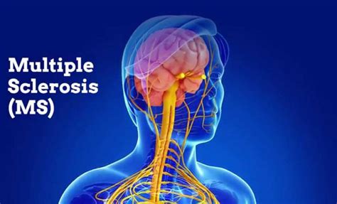
Epstein-Barr virus may be the leading cause of multiple sclerosis

Summary:

A new study provides compelling evidence of causality between Epstein-Barr virus and multiple sclerosis. It suggests that most MS cases could be prevented by stopping EBV infection, and that targeting EBV could lead to the discovery of a cure for MS.

Multiple sclerosis (MS), a progressive disease that affects 2.8 million people worldwide and for which there is no definitive cure, is likely caused by infection with the Epstein-Barr virus (EBV), according to a study led by Harvard T.H. Chan School of Public Health researchers.

Their findings will be published online in Science on January 13, 2022.

“The hypothesis that EBV causes MS has been investigated by our group and others for several years, but this is the first study providing compelling evidence of causality,” said Alberto Ascherio, professor of epidemiology and nutrition at Harvard Chan School and senior author of the study. “This is a big step because it suggests that most MS cases could be prevented by stopping EBV infection, and that targeting EBV could lead to the discovery of a cure for MS.”

MS is a chronic inflammatory disease of the central nervous system that attacks the myelin sheaths protecting neurons in the brain and spinal cord. Its cause is not known, yet one of the top suspects is EBV, a herpes virus that can cause infectious mononucleosis and establishes a latent, lifelong infection of the host. Establishing a causal relationship between the virus and the disease has been difficult because EBV infects approximately 95% of adults, MS is a relatively rare disease, and the onset of MS symptoms begins about ten years after EBV infection. To determine the connection between EBV and MS, the researchers conducted a study among more than 10 million young adults on active duty in the U.S. military and identified 955 who were diagnosed with MS during their period of service.

The team analyzed serum samples taken biennially by the military and determined the soldiers’ EBV status at time of first sample and the relationship between EBV infection and MS onset during the period of active duty. In this cohort, the risk of MS increased 32-fold after infection with EBV but was unchanged after infection with other viruses. Serum levels of neurofilament light chain, a biomarker of the nerve degeneration typical in MS, increased only after EBV infection. The findings cannot be explained by any known risk factor for MS and suggest EBV as the leading cause of MS.

Ascherio says that the delay between EBV infection and the onset of MS may be partially due the disease’s symptoms being undetected during the earliest stages and partially due to the evolving relationship between EBV and the host’s immune system, which is repeatedly stimulated whenever latent virus reactivates.

“Currently there is no way to effectively prevent or treat EBV infection, but an EBV vaccine or targeting the virus with EBV-specific antiviral drugs could ultimately prevent or cure MS,” said Ascherio.

Other Harvard Chan School researchers who contributed to this study include Kjetil Bjornevik, Marianna Cortese, Michael Mina, and Kassandra Munger.

Funding for this study came the National Institute of Neurological Disorders and Stroke, National Institutes of Health (NS046635, NS042194, and NS103891), the National Multiple Sclerosis Society (PP-1912-35234), the German Research Foundation (CO 2129/ 1-1), the National Institutes of Health (DP5- OD028145), and the Howard Hughes Medical Institute.


Story Source:

Materials provided by Harvard T.H. Chan School of Public Health. Note: Content may be edited for style and length.


Journal Reference:

  1. Kjetil Bjornevik, Marianna Cortese, Brian C. Healy, Jens Kuhle, Michael J. Mina, Yumei Leng, Stephen J. Elledge, David W. Niebuhr, Ann I. Scher, Kassandra L. Munger, Alberto Ascherio. Longitudinal analysis reveals high prevalence of Epstein-Barr virus associated with multiple sclerosis. Science, 2022 DOI: 10.1126/science.abj8222

  Harvard T.H. Chan School of Public Health. “Epstein-Barr virus may be leading cause of multiple sclerosis.” ScienceDaily. ScienceDaily, 13 January 2022. <www.sciencedaily.com/releases/2022/01/220113151342.htm>.


Let me introduce you to the concept of “neuritogenic” compounds: molecules that are known to regenerate nerve tissue and help repair damaged brains.

The Green Med Info website has a great database for published papers on different topics.

When using the search term “neuritogenic” some of the compounds that come up include:

  • Ubiquinol – CoQ 10
  • EGCG – as from green tea
  • Puerarin
  • Cannabinoids
  • Green Coffee Bean
  • Jujube
  • Lion’s Mane
  • Gensenosides
  • Curcumin

One key neuritogenic compound is sulforaphane with broccoli sprouts being the best source.

Part of the content for this article comes from an article published on the Natural News website


Further from the Natural News article:

Green Med Info has also published an overview of sulforaphane and its brain repair mechanisms as documented in published science. From that story:

The researchers determined the optimal concentration range of sulforaphane in promoting neural stem cell (NSC) growth without harming neurons. The researchers determined that “Concentrations of less than 5 mM did not induce cytotoxic e?ects, but rather potentially promote the growth of NSCs.”

Green Med Info has also published an overview of sulforaphane and its brain repair mechanisms as documented in published science. From that story:

The researchers determined the optimal concentration range of sulforaphane in promoting neural stem cell (NSC) growth without harming neurons. The researchers determined that “Concentrations of less than 5 mM did not induce cytotoxic e?ects, but rather potentially promote the growth of NSCs.” The term 5 mM means 5 milli-molar which is a reference to the concentration of sulforaphane in blood. 5 milli-molar is 5 thousands of a Mole. A Mole is a set number of molecules in one liter of a solution, regardless of molecular weight.

Although this depends a lot on body weight, we think it wouldn’t require much consumption of broccoli sprouts to achieve 5 mM concentrations in the blood of a person.

When neural stem cells were exposed to sulforaphane, they transformed into neurons

From the Green Med Info article:

…exposing NSCs to sulforaphane resulted in their differentiation [into] neurons, lending powerful support to the hypothesis that sulforaphane could stimulate brain repair.

This means that sulforaphane is a kind of molecular “activator” that causes neural stem cells to become neurons. This is how damaged brain cells are regrown.

How to grow your own sulforaphane for mere pennies

Because God and Mother Nature gave us all the medicine we need, you can “manufacture” your own sulforaphane for mere pennies and literally transform air into sulforaphane through the use of broccoli sprouts.

  1. Buy broccoli sprouts, a mason jar and a sprouting lid (see image below).
  2. Put about a tablespoon of broccoli spouting seeds into a mason jar, rinse with water, affix the sprouting lid and turn it upside to drain the water.
  3. Keep the sprouting jar upside down. Once a day, rinse with water and drain.
  4. In a few days, you have broccoli sprouts with loads of sulforaphane.

You can then eat the sprouts, use them in salads, blend them into smoothies, drop them into soups or whatever you want to do. Sulforaphane is a very robust molecule and it’s somewhat difficult to destroy, so don’t think you have to treat it like a delicate, fragile substance. Blending sprouts does not destroy their molecules. That’s because these molecules are very, very small.

Many grocery stores that offer sprouts typically also offer broccoli sprouts.

Other benefits of sulforaphane

Sulforaphane also exhibits powerful anti-cancer properties. GreenMedInfo lists the best properties of this molecule as reflected in published medical literature. It shows that sulforaphane is documented to help with:

  • oxidative stress
  • inflammation
  • prostate cancer
  • breast cancer
  • colon cancer
  • autism
  • pancreatic cancer
  • diabetes type-2
  • DNA damage
  • bladder cancer
  • insulin resistance

… and many other conditions.

Brain function is of course a key concern for many people these days and consuming foods which contain sulforaphane can help to maintain brain health.

I was reminded this morning of the significant potential health benefits of earthing/grounding while watching a program on GAIA.

If you are not familiar with GAIA, it is similar to Netflix but with more of an emphasis on topics such as integrative health, spirituality etc.

In fact I enjoy the content available on GAIA that I rarely watch Netflix any more.

Here is a link to the episode that I watched this morning on earthing/grounding which you can watch for free

Here is an example of what the published research has to say about the potential health benefits of earthing/grounding:
Environ Public Health. 2012; 2012: 291541. Published online 2012 Jan 12. doi: 10.1155/2012/291541 PMCID: PMC3265077PMID: 22291721

Earthing: Health Implications of Reconnecting the Human Body to the Earth’s Surface Electrons

Gaétan Chevalier, 1, 2 ,*Stephen T. Sinatra, 3 James L. Oschman, 4 Karol Sokal, 5 and Pawel Sokal 6

Abstract

Environmental medicine generally addresses environmental factors with a negative impact on human health. However, emerging scientific research has revealed a surprisingly positive and overlooked environmental factor on health: direct physical contact with the vast supply of electrons on the surface of the Earth.

Modern lifestyle separates humans from such contact. The research suggests that this disconnect may be a major contributor to physiological dysfunction and unwellness. Reconnection with the Earth’s electrons has been found to promote intriguing physiological changes and subjective reports of well-being. Earthing (or grounding) refers to the discovery of benefits—including better sleep and reduced pain—from walking barefoot outside or sitting, working, or sleeping indoors connected to conductive systems that transfer the Earth’s electrons from the ground into the body. This paper reviews the earthing research and the potential of earthing as a simple and easily accessed global modality of significant clinical importance.

2.1. Sleep and Chronic Pain

2.3. Earthing Reduces Electric Fields Induced on the Body

2.4. Physiological and Electrophysiological Effects

4.4. Heart Rate Variability

4.5. Reduction of Primary Indicators of Osteoporosis, Improvement of Glucose Regulation, and Immune Response

4.6. Altered Blood Electrodynamics

Table 1

Subjective sleep, pain, and well-being feedback.

CategoriesTest subjects*Control subjects**
SameImprovedSameImproved
Time to fall asleep4 = 15%23 = 85%20 = 87%3 = 13%
Quality of sleep2 = 7%25 = 93%20 = 87%3 = 13%
Wake feeling rested0 = 0%27 = 100%20 = 87%3 = 13%
Muscles stiffness and pain5 = 18%22 = 82%23 = 100%0 = 0%
Chronic back and/or joint pain7 = 26%20 = 74%23 = 100%0 = 0%
General well-being6 = 22%21 = 78%20 = 87%3 = 13%

*Reports not received from three participants.

**Reports not received from seven participants.

4. Conclusion

De Flora et al. wrote the following: “Since the late 20th century, chronic degenerative diseases have overcome infectious disease as the major causes of death in the 21st century, so an increase in human longevity will depend on finding an intervention that inhibits the development of these diseases and slows their progress” [33].

Could such an intervention be located right beneath our feet? Earthing research, observations, and related theories raise an intriguing possibility about the Earth’s surface electrons as an untapped health resource—the Earth as a “global treatment table.” Emerging evidence shows that contact with the Earth—whether being outside barefoot or indoors connected to grounded conductive systems—may be a simple, natural, and yet profoundly effective environmental strategy against chronic stress, ANS dysfunction, inflammation, pain, poor sleep, disturbed HRV, hypercoagulable blood, and many common health disorders, including cardiovascular disease. The research done to date supports the concept that grounding or earthing the human body may be an essential element in the health equation along with sunshine, clean air and water, nutritious food, and physical activity.

This simple behavioral change has the potential to initiate powerful improvements in health conditions and is something that may warrant consideration for everyone.