Today I want to share with you an article from the Hormones Matter website written by Chandler Marrs, PhD
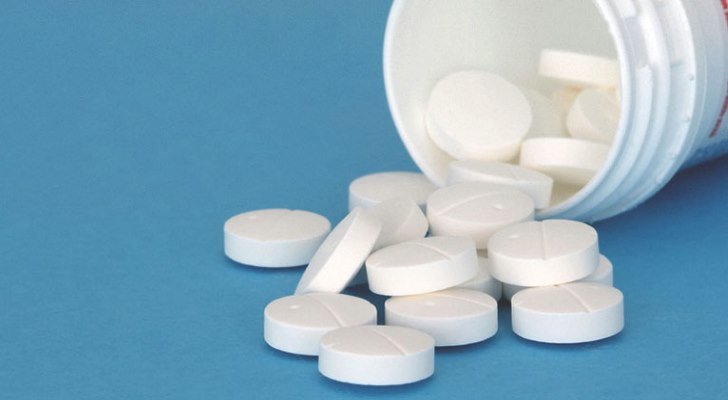
The article focuses on the fact that many individuals are consuming Metformin considering it to be a magical anti-aging drug.
I am in agreement with Chandra in that personally I have have never been a fan.
There are several considerations for myself as to why I feel this way which she talks about in this article, such as deficiencies that can develop, negative effects on mitochondrial function and a potential negative impact on exercise performance.
I would suggest that berberine provides many of the same benefits as Metformin as well as some such as CV benefits that Metformin does not provide – and berberine does not have any of the same negative effects vs. Metformin.
Following is Chandler’s article
I have never been a fan of Metformin. It seemed too good to be true. Many years ago I had a conversation with a researcher about all of its possible therapeutic indications. His lab was actively pursuing the anti-cancer angle. That should have been a clue that Metformin might be causing more damage than we recognized, but it wasn’t. At that point, I was still enamored with the wonders of pharmacology and hadn’t yet begun my path toward understanding medication adverse reactions. Indeed, it wasn’t until very recently, when a family member began suffering from one of these reactions, that I began my investigation in full. This is what I learned.
This article was based upon a published study done at Washington State University and published in The Lancet Diabetes & Endocrinology – the citation is included at the end of this article.
” Due to its phenolic structure BPA has been shown to interact with estrogen receptors and to act as agonist or antagonist via estrogen receptor (ER) dependent signalling pathways. Therefore, BPA has been shown to play a role in the pathogenesis of several endocrine disorders including female and male infertility, precocious puberty, hormone dependent tumours such as breast and prostate cancer and several metabolic disorders including polycystic ovary syndrome (PCOS)”
Due to the prevalence of exposure to BPA in our environment – as well as other chemicals and heavy metals periodic monitoring and supervised detox programs to clear out this toxin load are serious considerations for optimizing health and potentially extending healthspan.
Summary:
Researchers have developed a more accurate method of measuring bisphenol A (BPA) levels in humans and found that exposure to the endocrine-disrupting chemical is far higher than previously assumed. The study provides the first evidence that the measurements relied upon by regulatory agencies, including the US Food and Drug Administration, are flawed, underestimating exposure levels by as much as 44 times. Researchers have developed a more accurate method of measuring bisphenol A (BPA) levels in humans and found that exposure to the endocrine-disrupting chemical is far higher than previously assumed.
The study, published in the journal The Lancet Diabetes & Endocrinology
on Dec. 5, provides the first evidence that the measurements relied
upon by regulatory agencies, including the U.S. Food and Drug
Administration, are flawed, underestimating exposure levels by as much
as 44 times.
“This study raises serious concerns about
whether we’ve been careful enough about the safety of this chemical,”
said Patricia Hunt, Washington State University professor and
corresponding author on the paper. “What it comes down to is that the
conclusions federal agencies have come to about how to regulate BPA may
have been based on inaccurate measurements.”
BPA can be found in a wide range of
plastics, including food and drink containers, and animal studies have
shown that it can interfere with the body’s hormones. In particular,
fetal exposure to BPA has been linked to problems with growth,
metabolism, behavior, fertility and even greater cancer risk.
Despite this experimental evidence, the
FDA has evaluated data from studies measuring BPA in human urine and
determined that human exposure to the chemical is at very low, and
therefore, safe levels. This paper challenges that assumption and raises
questions about other chemicals, including BPA replacements, that are
also assessed using indirect methods.
Hunt’s colleague, Roy Gerona, assistant
professor at University of California, San Francisco, developed a direct
way of measuring BPA that more accurately accounts for BPA metabolites,
the compounds that are created as the chemical passes through the human
body.
Previously, most studies had to rely on
an indirect process to measure BPA metabolites, using an enzyme solution
made from a snail to transform the metabolites back into whole BPA,
which could then be measured.
Gerona’s new method is able to directly measure the BPA metabolites themselves without using the enzyme solution.
In this study, a research team comprised
of Gerona, Hunt and Fredrick vom Saal of University of Missouri compared
the two methods, first with synthetic urine spiked with BPA and then
with 39 human samples. They found much higher levels of BPA using the
direct method, as much as 44 times the mean reported by the National
Health and Nutrition Examination Survey (NHANES). The disparity between
the two methods increased with more BPA exposure: the greater the
exposure the more the previous method missed.
Gerona, the first author on the paper, said more replication is needed.
“I hope this study will bring attention
to the methodology used to measure BPA, and that other experts and labs
will take a closer look at and assess independently what is happening,”
he said.
The research team is conducting further
experiments into BPA measurement as well as other chemicals that may
also have been measured in this manner, a category that includes
environmental phenols such as parabens, benzophenone, triclosan found in
some cosmetics and soaps, and phthalates found in many consumer
products including toys, food packaging and personal care products.
“BPA is still being measured indirectly
through NHANES, and it’s not the only endocrine-disrupting chemical
being measured this way,” Gerona said. “Our hypothesis now is that if
this is true for BPA, it could be true for all the other chemicals that
are measured indirectly.”
This study was supported by grants from the National Institutes of Health.
Roy Gerona, Frederick S vom Saal, Patricia A Hunt. BPA: have flawed analytical techniques compromised risk assessments?The Lancet Diabetes & Endocrinology, 2019; DOI: 10.1016/S2213-8587(19)30381-X
Bisphenol A (BPA) belongs to chemicals
that are produced in large quantities worldwide. It is commonly used as
monomer in polycarbonate synthesis, plasticizer in the production of
epoxy resins, as well as an additive for the elimination of surfeit of
hydrochloric acid during the polyvinyl chloride (PVC) production. BPA is
not only used in the production of plastics intended to a direct
contact with food, including plastic packaging and kitchenware, but also
in inner coatings of cans and jar caps. There are various routes of
human exposure to this substance such as oral, by inhalation and
transdermal. The main sources of exposure to BPA include food packaging
and dust, dental materials, healthcare equipment, thermal paper, toys
and articles for children and infants. BPA is metabolized in the liver
to form bisphenol A glucuronide and mostly in this form is excreted with
urine. Due to its phenolic structure BPA has been shown to interact
with estrogen receptors and to act as agonist or antagonist via estrogen
receptor (ER) dependent signalling pathways. Therefore, BPA has been
shown to play a role in the pathogenesis of several endocrine disorders
including female and male infertility, precocious puberty, hormone
dependent tumours such as breast and prostate cancer and several
metabolic disorders including polycystic ovary syndrome (PCOS).
Because of the constant, daily exposure and its tendency to
bio-accumulation, BPA seems to require special attention such as
biomonitoring. This observation should include clinical tests of BPA
concentration in the urine, which is not only one of the best methods of
evaluation of the exposure to this compound, but also the dependence of
the daily intake of BPA and the risk of some endocrine disorders.
PMID: 25813067
People who live with depression have low blood levels of a specific molecule, new medical research has revealed. It’s called acetyl-L-carnitine, and those with particularly severe, treatment-resistant or childhood onset depression were found to have the lowest levels.
Naturally produced by the body, acetyl-L-carnitine plays a crucial
role in metabolising fat and the production of energy. It’s also widely
available as a dietary supplement – not some strange and esoteric thing.
Now
researchers from multiple institutions have found a link to depression,
noticing a clear correlation between the condition and noticeably low
levels of acetyl-L-carnitine.
In recent years, more and more
evidence has been building to suggest this link. Since at least 1991,
medical researchers have been aware of acetyl-L-carnitine’s potential to
treat depression, particularly in geriatric and comorbid patients, with the substance showing greater efficacy than a placebo.
More recently, Carla Nasca of the Rockefeller University led a study on rodents,
which found that acetyl-L-carnitine had a fast-acting antidepressant
effect on rats, kicking into effect in just a few days, rather than the
weeks it takes for drugs like SSRIs.
Now Nasca and colleagues have conducted a study on human patients to see if there’s a basis for a similar trial in people.
“It’s the number one reason for absenteeism at work, and one of
the leading causes of suicide. Worse, current pharmacological
treatments are effective for only about 50 percent of the people for
whom they’re prescribed. And they have numerous side effects, often
decreasing long term compliance.”
The research team recruited 71
patients with a diagnosis of depression. These were men and women, aged
between 20 and 70. They also recruited 45 demographically matched
healthy controls.
The patients had to fill out a detailed
questionnaire, undergo a clinical assessment and medical history, and
give a blood sample. Of the patients with depression, 28 had moderate
depression and 43 had severe depression at the time of the study.
When
compared to the age- and sex-matched healthy controls, the patients
with depression had substantially lower levels of acetyl-L-carnitine.
Those
with the most severe depression had the lowest levels. This included
patients whose depression had resisted antidepressant drugs, those with
early onset, and those who had experienced childhood abuse, neglect,
poverty or violence.
These patients constitute around 25-30
percent of all people suffering depression, and are the most in need of
help, the researchers said.
But there are a few steps to be done before acetyl-L-carnitine
supplements can be approved as a treatment. In particular, clinical
trials on human patients with depression, since, as we know, results from rodent models can’t always be replicated in humans.
The
researchers also don’t know the reason for the correlation, or the
effect it has. The rat research suggests that acetyl-L-carnitine plays a
role in the brain, preventing the excessive firing of excitatory
neurons, but this will need to be explored further as well.
“We’ve identified an important new biomarker of major depression disorder,” Rasgon said.
“We
didn’t test whether supplementing with that substance could actually
improve patients’ symptoms. What’s the appropriate dose, frequency,
duration? We need to answer many questions before proceeding with
recommendations, yet. This is the first step toward developing that
knowledge, which will require large-scale, carefully controlled clinical
trials.”
And we’ll be eagerly awaiting the results of those trials.
Meanwhile, the team’s research can be found in the journal PNAS.
Dietary intervention restores protective protein and decreases death rate in mice
Source: Society for Neuroscience
The incidence of dementia and Alzheimer’s continues to escalate in the general population.
LCHF/Keto diets have proven to be beneficial to individuals dealing with these health issues.
It has been suggested that these conditions may partly be due to impaired glucose metabolism in the brain, hence the increasing use of the term “Type 3 Diabetes”.
Enabling the brain to use ketones for its energy source therefore can provide some benefit with regards to brain function.
A major challenge with this is that a radical dietary shift in the geriatric population can be quite challenging – if not impossible.
Usage of exogenous ketone compounds is one potential option in this situation.
Following is an article from Science Daily which talks about published research which suggests that increasing ketone levels in the diet can help to protect neurons from death during the progression of Alzheimer’s disease.
Summary: A ketone-supplemented diet may protect neurons from death during the progression of Alzheimer’s disease, according to research in mice.
A
ketone-supplemented diet may protect neurons from death during the
progression of Alzheimer’s disease, according to research in mice
recently published in JNeurosci.
Early in the development of Alzheimer’s
disease, the brain becomes over excited, potentially through the loss of
inhibitory, or GABAergic, interneurons that keep other neurons from
signaling too much. Because interneurons require more energy compared to
other neurons, they may be more susceptible to dying when they
encounter the Alzheimer’s disease protein amyloid beta. Amyloid beta has
been shown to damage mitochondria — the metabolic engine for cells —
by interfering with SIRT3, a protein that preserves mitochondrial
functions and protects neurons.
Cheng et al. genetically reduced levels
of SIRT3 in mouse models of Alzheimer’s disease. Mice with low levels of
SIRT3 experienced a much higher mortality rate, more violent seizures,
and increased interneuron death compared to the mice from the standard
Alzheimer’s disease model and control mice. However, the mice with
reduced levels of SIRT3 experienced fewer seizures and were less likely
to die when they ate a diet rich in ketones, a specific type of fatty
acid. The diet also increased levels of SIRT3 in the mice.
Increasing SIRT3 levels via ketone
consumption may be a way to protect interneurons and delay the
progression of Alzheimer’s disease.
Story Source:
Materials provided by Society for Neuroscience. Note: Content may be edited for style and length.
Journal Reference:
Aiwu Cheng, Jing Wang, Nathaniel Ghena,
Qijin Zhao, Isabella Perone, M. Todd King, Richard L. Veech, Myriam
Gorospe, Ruiqian Wan, Mark P. Mattson. SIRT3 Haploinsufficiency
Aggravates Loss of GABAergic Interneurons and Neuronal Network
Hyperexcitability in an Alzheimer’s Disease Model. The Journal of Neuroscience, 2019; 1446-19 DOI: 10.1523/JNEUROSCI.1446-19.2019
Abstract
SIRT3 Haploinsufficiency
Aggravates Loss of GABAergic Interneurons and Neuronal Network
Hyperexcitability in an Alzheimer’s Disease Model
Impaired mitochondrial function and
aberrant neuronal network activity are believed to be early events in
the pathogenesis of Alzheimer’s disease (AD), but how mitochondrial
alterations contribute to aberrant activity in neuronal circuits is
unknown. In this study, we examined the function of mitochondrial
protein deacetylase sirtuin 3 (SIRT3) in the pathogenesis of AD.
Compared to AppPs1 mice, Sirt3-haploinsufficient AppPs1 mice
(Sirt3+/-AppPs1) exhibit early epileptiform EEG activity and Seizure.
Both male and female Sirt3+/-AppPs1 mice were observed to die
prematurely before five months of age.
When comparing male mice among different genotypes, Sirt3
haploinsufficiency renders GABAergic interneurons in the cerebral cortex
vulnerable to degeneration and associated neuronal network
hyperexcitability. Feeding Sirt3+/-AppPs1 AD mice with a ketone
ester-rich diet increases SIRT3 expression and prevents seizure-related
death and the degeneration of GABAergic neurons, indicating that the
aggravated GABAergic neuron loss and neuronal network hyperexcitability
in Sirt3+/-AppPs1 mice are caused by SIRT3 reduction and can be rescued
by increase of SIRT3 expression. Consistent with a protective role in
AD, SIRT3 levels are reduced in association with cerebral cortical Aβ
pathology in AD patients. In summary, SIRT3 preserves GABAergic
interneurons and protects cerebral circuits against hyperexcitability,
and this neuroprotective mechanism can be bolstered by dietary ketone
esters.
SIGNIFICANCE STATEMENT
GABAergic neurons provide the main
inhibitory control of neuronal activity in the brain. By preserving
mitochondrial function, SIRT3 protects parvalbumin and calretinin
interneurons against Aβ-associated dysfunction and degeneration in
AppPs1 AD mice, thus restraining neuronal network hyperactivity. The
neuronal network dysfunction that occurs in AD can be partially reversed
by physiological, dietary, and pharmacological interventions to
increase SIRT3 expression and enhance the functionality of GABAergic
interneurons.
Fasting in its many forms can provide profound beneficial health benefits.
Following is an article on this topic authored by Dr. Dan Pompa which provides a good overview.
Regards,
Robert (Rob) Lamberton
Fasting is a very old ritual to boost health that is found in religions all over the world and is rooted in natural ancestral cycles of feast and famine. Before we had grocery stores, restaurants, and even food delivery services- there were often times with very little to no food. Following times of famine, there was an abundance of food (following a successful harvest, forage, or hunt). Even animal wisdom harnesses the power of fasting- like dogs, that will intuitively stop eating when they are sick. More and more studies are emerging on the incredible benefits that fasting can have, on not only for health but also suggesting a boost in longevity.
Fasting diets
have nothing to do with WHAT or HOW MUCH you eat, but WHEN you eat.
Intermittent fasting (or IF) is the art of restricted time eating, so instead
of counting calories or restricting what types of foods you eat- the entire
“diet” relies on when you do, and don’t eat.
Recent Research on Fasting
Have Your Cake And Eat It Too: Boost Health
and Longevity Not By Changing What You Eat, But When You
Eat.
Intermittent Fasting Research
Although Intermittent Fasting to boost health has gained
popularity in more recent years, its wisdom dates back to our ancestors from
the stone age. Apart from periods of feast and famine, our ancestors’ lives
were also heavily dictated by the rising and setting of the sun; activities
like eating naturally happened during day time. Our exposure to light, food,
and movement are the main tenets that inform and program our circadian rhythm.
This internal rhythm influences everything from sleep-wake cycles, hormone
release, eating habits and digestion, body temperature, and other important
bodily functions.1 Intermittent fasting plays a role in giving the
body an adequate period of rest from digestion, enabling it to not only heal-
but thrive.
Research on Fasting is Extensive
Many of the
studies regarding fasting to boost health and longevity have been done on
animals. However, these studies suggest promising effects on metabolic
functions, health, and lifespan for humans. Although there are many variables,
Rafael deCabo, a scientist at the National Institute on Aging and the
study’s lead author explains that;
“in the absence of
calorie restriction, and independent of diet composition, fasting mice do
better than non-fasting”.2
Boost Health! The ever-increasing research
regarding fasting suggests some incredible health and longevity benefits
including:
Autophagy
A boost in stem cells
Boost in ketones
Hormone optimization
Increased insulin sensitivity
Reset of the microbiome
Reset of the DNA (gene code)
Decrease in inflammation
A decrease in oxidative stress
Reduced instances of chronic disease and obesity
Protection against unusual deterioration of cognitive function
Fat loss
Cancer prevention
Promotion of better sleep
More satiety/ reduced hunger
Although benefits
are often examined as individual points, they are in fact very much intertwined
to promote overall longevity. One of the main ways IF leads to longevity is
“multi-system regeneration,” which fasting researcher Dr. Valter Longo explains
occurs during the presence of ketones in the blood. The autophagy process that
happens during a fasting period breaks down weak and damaged cells, which are
then replaced with new stem cells after food is reintroduced.
“You get rid of
the junk during starvation — and once you have food, you can rebuild… The
damaged cells are replaced with new cells, working cells — and now the system
starts working properly.”
Research on Fasting: Health and Longevity
All these
benefits suggest a direct link between fasting and longevity, although
conducting a clinical longevity study in humans is unfeasible at the moment,
for would cost “a hundred million dollars or more,” according to Longo. “But if
you look at the data from our trial … it would be hard to see how they would
not live longer.”
Dr. Valter Longo
and Dr. Satchin Panda’s study demonstrated that a 12-hour feeding window
reduced blood cholesterol, fasting blood sugar, body weight, body fat,
inflammation, and dysbiosis, and increased energy expenditure, motor control,
endurance, sleep, and cardiac function.3 Their study examined the
intricate relationship between time-restricted feeding (IF), circadian health,
and ultimately concluded that simply limiting your eating window to a minimum
of 12 hours reduces biological age irrelevant of any dietary changes! Indeed,
their study suggests that you can have your cake and eat it too… so long as you
do so within your eating window.
Research on Fasting: How To Do It
There are many
different fasting styles that range from multiple days water-only fasts, to
bone broth fasts, to alternate day fasting… but intermittent fasting itself is
conceptually incredibly simple: engage in a particular restricted eating
window, preferably rooted in 2 meals (and no snacking). This might seem not too
far off from your current habits, but studies show the average American eats
17-21 times a day! This is detrimental to our health and longevity.
Classic Intermittent Fasting: The Eating
Window
The key is,
aforementioned, restricting your eating window. The science suggests a very minimum
of 12 hours to see any benefits, so if you have no experience fasting- start
there. If you eat your first meal at 8 am, no food (or beverage other than
plain water) after 8 pm.4 From there, extend the fasting window to
ideally (at least) 16 hours. Whether you decide to skip breakfast or dinner is
completely personal, find what works best for your schedule and which option is
more sustainable over the long run. A 2018 study comparing a 12-hour feeding
window to an 8-hour feeding window demonstrated that although both groups lost
weight, those in the 8-hour feeding window group dramatically lower insulin
levels, improved insulin sensitivity, and significantly lower blood pressure in
only five weeks.5
Research on Fasting: One Meal a Day
“One meal a day”
(or OMAD) is an extreme version of intermittent fasting. An individual shortens
their eating window to essentially the duration of one single meal. The
benefits of this technique essentially amplify all the aforementioned benefits
of a 16/8 IF protocol. OMAD gives the body even more time in this resting
(vs. digesting) state. OMAD is not, however, for everyone- nor should it be the
goal. Consuming one meal a day can be more taxing on the adrenal system. OMAD
could even induce more detoxification than an individual can handle at once.
Like any type of
good stress (exercise, sauna, cold therapy), the adrenals and overall system
need to be strong enough to withstand the short term stressor. Ease into
intermittent fasting at your own pace, and always listen to your body. A great
way to transition into it and/ or reboot your system is to take part in the
5-day Fasting Mimicking Diet™.
Research on Fasting to Boost Health and
Longevity: The Fasting Mimicking DietTM
Fasting for health and longevity can be a daunting endeavor for someone who is used to eating 3+ meals a day their entire lives, and this is where the fasting mimicking diet comes in. Fasting expert and researcher Dr. Valter Longo created the Fasting Mimicking Diet program that mimics the benefits of a fasting protocol, combining both the benefits of intermittent fasting and a longer term fast (through caloric restriction). Prolon® takes out the guesswork but providing clients with all their meals for a 5 day period. Longo is the Director of both the Longevity Institute at the University of Southern California and The Program on Longevity and Cancer at IFOM in Milan, and his clinical study demonstrated remarkable benefits that fasting has to offer in just 5 days (repeated for 3 months):
Promote stem cell-based renewal in the body
Decrease excess body fat while preserving lean muscle mass
Maintain healthy levels of blood glucose, cholesterol, & blood pressure
Decreased hormone IGF-1 (which has been implicated with aging and disease)6
We suggest using
this fasting
mimicking diet to boost health if you are completely new to fasting
or are trying to break destructive eating patterns! This can be a bridge to
continue on with regular Intermittent Fasting thereafter!
References
Longo, Valter D., and Satchidananda Panda. “Fasting, Circadian Rhythms, and Time-Restricted Feeding in Healthy Lifespan.” Cell Metabolism, vol. 23, no. 6, 2016, pp. 1048–1059., doi:10.1016/j.cmet.2016.06.001.
Mitchell, Sarah J., et al. “Daily Fasting Improves Health and Survival in Male Mice Independent of Diet Composition and Calories.” Cell Metabolism, vol. 29, no. 1, Jan. 2019, doi:10.1016/j.cmet.2018.08.011
Sutton, Elizabeth F., et al. “Early Time-Restricted Feeding Improves Insulin Sensitivity, Blood Pressure, and Oxidative Stress Even without Weight Loss in Men with Prediabetes.” Cell Metabolism, vol. 27, no. 6, 2018, doi:10.1016/j.cmet.2018.04.010.
Wei, Min, et al. “Fasting-Mimicking Diet and Markers/Risk Factors for Aging, Diabetes, Cancer, and Cardiovascular Disease.” Science Translational Medicine, vol. 9, no. 377, 2017, doi:10.1126/scitranslmed.aai8700.
We love sweet treats. But too much sugar in our diets can lead to weight gain and obesity, Type 2 diabetes and dental decay. We know we shouldn’t be eating candy, ice cream, cookies, cakes and drinking sugary sodas, but sometimes they are so hard to resist.
It’s as if our brain is hardwired to want these foods.
As a neuroscientist my research centres on how modern day “obesogenic”, or obesity-promoting, diets
change the brain. I want to understand how what we eat alters our
behaviour and whether brain changes can be mitigated by other lifestyle
factors.
Your body runs on sugar – glucose to be precise. Glucose comes from the Greek word glukos which means sweet. Glucose fuels the cells that make up our body – including brain cells (neurons).
Dopamine “hits” from eating sugar
On
an evolutionary basis, our primitive ancestors were scavengers. Sugary
foods are excellent sources of energy, so we have evolved to find sweet
foods particularly pleasurable. Foods with unpleasant, bitter and sour
tastes can be unripe, poisonous or rotting – causing sickness.
So
to maximize our survival as a species, we have an innate brain system
that makes us like sweet foods since they’re a great source of energy to
fuel our bodies.
When we eat sweet foods the brain’s reward system – called the mesolimbic dopamine system – gets activated. Dopamine
is a brain chemical released by neurons and can signal that an event
was positive. When the reward system fires, it reinforces behaviours –
making it more likely for us to carry out these actions again.
Dopamine “hits” from eating sugar promote rapid learning to preferentially find more of these foods.
Our
environment today is abundant with sweet, energy rich foods. We no
longer have to forage for these special sugary foods – they are
available everywhere.
Unfortunately, our brain is still
functionally very similar to our ancestors, and it really likes sugar.
So what happens in the brain when we excessively consume sugar?
Can sugar rewire the brain?
The brain continuously remodels and rewires itself through a process called neuroplasticity.
This rewiring can happen in the reward system. Repeated activation of
the reward pathway by drugs or by eating lots of sugary foods causes the
brain to adapt to frequent stimulation, leading to a sort of tolerance.
In the case of sweet foods, this means we need to eat more to get the same rewarding feeling – a classic feature of addiction.
Food addiction
is a controversial subject among scientists and clinicians. While it is
true that you can become physically dependent on certain drugs, it is
debated whether you can be addicted to food when you need it for basic survival.
The brain wants sugar, then more sugar
Regardless of
our need for food to power our bodies, many people experience food
cravings, particularly when stressed, hungry or just faced with an
alluring display of cakes in a coffee shop.
To resist cravings, we
need to inhibit our natural response to indulge in these tasty foods. A
network of inhibitory neurons is critical for controlling behaviour.
These neurons are concentrated in the prefrontal cortex – a key area of the brain involved in decision-making, impulse control and delaying gratification.
Importantly,
this shows that what we eat can influence our ability to resist
temptations and may underlie why diet changes are so difficult for
people.
Importantly,
the brain’s neuroplasticity capabilities allow it to reset to an extent
following cutting down on dietary sugar, and physical exercise can augment this process.
Foods rich in omaga-3 fats (found in fish oil, nuts and seeds) are also
neuroprotective and can boost brain chemicals needed to form new
neurons.
While it’s not easy to break habits like always eating
dessert or making your coffee a double-double, your brain will thank you
for making positive steps.
The first step is often the hardest. These diet changes can often get easier along the way.